
- Mucocele is a common lesion of the oral mucosa.
- It’s a clinical term (based on the location of the lesion, clinical appearance, history given by patient etc.).
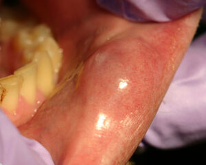
Mucocele on Lower Lip
Histo-pathologically Mucocele Is Of Two Types:-
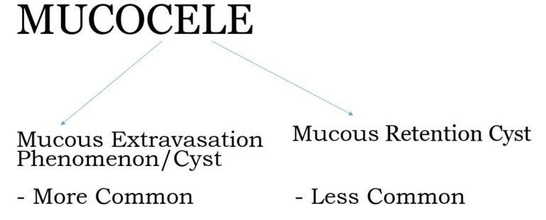
Mucous Extravasation Cyst
- Lesions in which mucus has extravasated into connective tissue from a severed excretory duct.
- Mainly affect the minor salivary glands, particularly of the lip.
- Not a TRUE CYST (no epithelial lining).
How Is A Mucous Extravasation Cyst Formed?
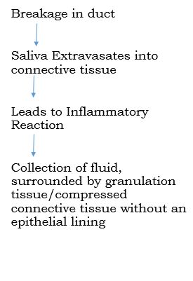
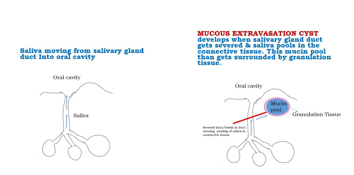
Normal Salivary Flow Phenomenon Vs How A Mucous Extravasation Cyst Is Formed
Clinical Features
- Most common site is lower lip (as a result of injury or biting of mucosa).
- Other sites:
- Buccal mucosa
- Floor of the mouth
- Ventral tongue
- Soft palate
- Retromolar area
- Upper lip is an uncommon site
- Age – Children & young adults
- Sex Predilection – Male = Female
- Usually superficial
- Size – Rarely larger than 1 cm in diameter
- Appearance – Dome-shaped swellings, fluctuant, translucent bluish due to thin wall
- Duration – Variable, Characteristic Finding: Alternate regression and recurrence due to the cystic cavity getting rupture and re-accumulation of saliva. Post rupture, they create painful ulcerations which heal within days.
- Sometimes Superficial Mucocele (Variant) – Occur on soft palate, retromolar area, and posterior buccal mucosa and they appear as 1-4 mm tense vesicles that burst leaving behind painful ulcers. This should not be confused with vesiculo-bullous lesions.
- A large mucocele in the floor of the mouth is called a Ranula
Histopathologic Features
Microscopically there is:-
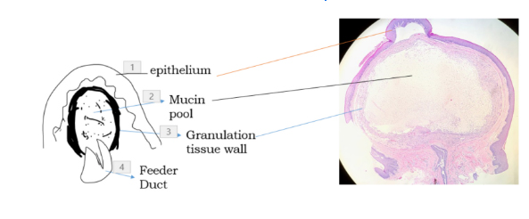
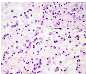
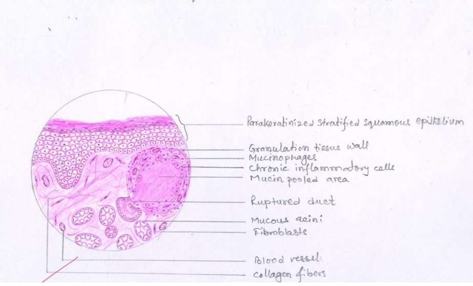
- Mucin Pool
- Surrounded by Granulation Tissue Wall /compressed connective tissue wall in long standing lesions
- No epithelial lining
- Prominent cells Mucinophages – Macrophages migrate into the mucin pool and by phagocytosing it develop foamy or vacuolated cytoplasm
- Sometimes – Ruptured salivary duct feeding into the area (called as Feeder Duct)
- Adjacent minor salivary glands often contain chronic inflammatory cell infiltrate & dilated ducts
Treatment & Prognosis
- Short-lived lesions rupture and heal by themselves
- Long Standing – Surgical Excision along with adjacent minor salivary gland tissue
- Prognosis is excellent, although sometimes may recur, especially if feeding glands not removed
Mucous Retention Cyst
- Swelling due to obstruction of salivary gland excretory duct
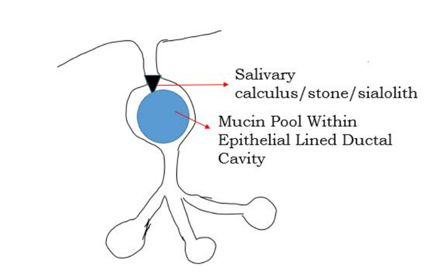
- Obstruction – salivary calculus/sialolith/salivary stone. Sometimes caused by periductal scarring or an impinging tumor
- Accumulation of mucin within epithelial lined ductal cavity i.e. ductal dilatation secondary to obstruction
- TRUE CYST – a retention cyst is lined by flattened/compressed ductal epithelium
- Less common than mucous extravasation cyst
Clinical Features
- Clinically difficult to differentiate from mucous extravasation cyst
- Affects adults
- SITE
1. Major or minor salivary gland
2. Intraoral Cysts commonly involve minor salivary glands of floor of mouth, buccal mucosa, lips - Pain especially at meal time
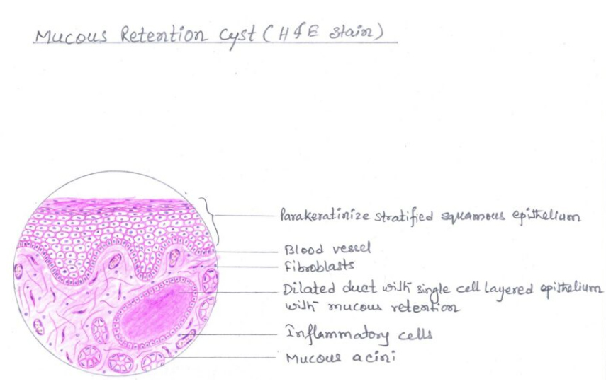
Histopathologic Features
- Epithelial lining is variable
- May consist of cuboidal , columnar, or atrophic squamous epithelium surrounding thin or mucoid secretions in the lumen
- The epithelium may undergo oncocytic metaplasia appearing as papillary folds into the cystic lumen

Treatment
- Surgical Excision along with adjacent minor salivary gland tissue
References
- Shafer’s Textbook Of Oral Pathology
- Shear – Cysts Of The Oral & Maxillofacial Regions
- Neville – Oral & Maxillofacial Pathology
- Image – Wikipedia & Wikimedia Commons


