

OUTLINE
- INTRODUCTION
- DEFINITION
- ORIGIN
- CLINICAL FEATURES
- RADIOGRAPHIC FEATURES
- HISTOPATHOLOGIC FEATURES
- TREATMENT & PROGNOSIS
INTRODUCTION
- Odontoma
- odont means of odontogenic origin
- oma means benign tumor
- Thus by definition alone, Odontoma refers to any tumor of odontogenic origin
- Reported to be the most common of all odontogenic neoplasms and tumour-like lesions
- Many authorities believe odontomas to be developmental anomalies (hamartomas), rather than true neoplasms
DEFINITION
"a growth in which both the epithelial and the mesenchymal cells exhibit complete differentiation, with the result that functional ameloblasts and odontoblasts form enamel and dentin. This enamel and dentin are usually laid down in an abnormal pattern because the organization of the odontogenic cells fails to reach a normal state of morphodifferentiation"
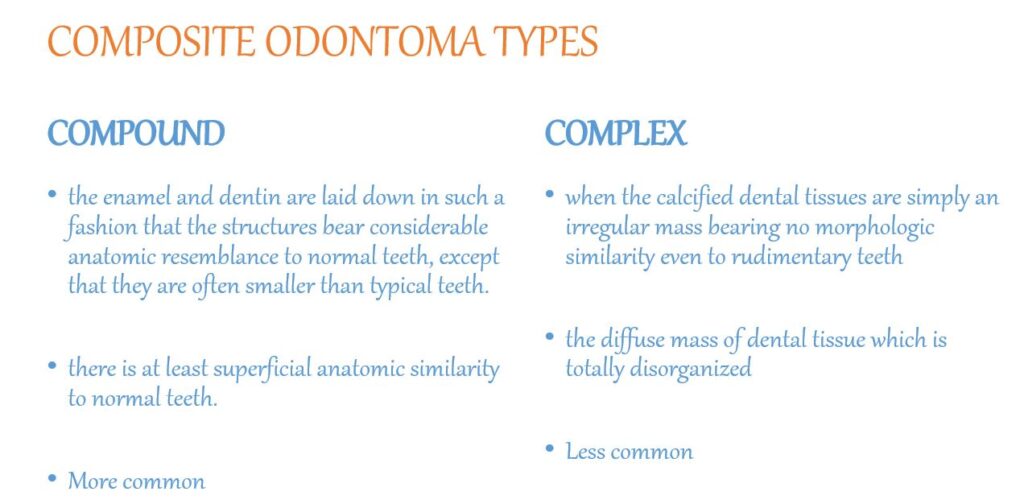
This lesion is composed of more than one type of tissue therefore called a COMPOSITE ODONTOMA
- COMPOSITE ODONTOMA is of 2 Types:-
- Compound Composite Odontoma
- Complex Composite Odontoma
ETIOLOGY
- The extraneous bud of the dental lamina is considered to be the tissue of origin for odontoma
- Exact cause unknown. Possible causes:-
- local trauma
- infection
- either inherited or are due to a mutant gene or interference, possibly postnatal, with the genetic control of tooth development
CLINICAL FEATURES
- AGE –
- any age
- mean age 14.8 years
- common in second decade of life
- SEX PREDILECTION –
- no gender predilection

- Slight male predilection (some studies)
- no gender predilection
- SITE –
- 67% in maxilla and 33% in mandible
- Compound odontoma more common than Complex odontoma
- Compound Odontoma – common in the anterior maxilla (incisor, canine area)
- Complex Odontoma – common in posterior mandible (1st & 2nd molar area followed by premolar area)
- Both types – common on right side of the jaw than on the left
- Generally asymptomatic
- Occasionally signs and symptoms relating to their presence do occur. These generally consist of:-
- unerupted or impacted teeth
- retained deciduous teeth
- swelling & evidence of infection
- Usually remains small, the diameter of the mass only occasionally greatly exceeding that of a tooth. Occasionally, it does become large and may produce expansion of bone with consequent facial asymmetry
- Sometimes they erupt also
RADIOGRAPHIC FEATURES
- Characteristic
- Since most odontomas are clinically asymptomatic and are discovered by routine radio-graphic examination, the dentist should be familiar with their appearance
- Often situated between the roots of teeth
- Compound Odontoma – Variable number of tooth like structures with the same peripheral outline. It may contain only a few structures resembling teeth, or it may contain several dozen
Or
Complex Odontoma – Irregular mass of calcified material surrounded by a narrow radiolucent band with a smooth outer periphery
- Both forms of odontoma are frequently associated with unerupted teeth
- Complex odontoma may appear also as a calcified mass overlying the crown of an unerupted or impacted tooth
- A developing odontoma may be discovered on the routine radiograph and present difficulty in diagnosis because of the lack of calcification
HISTOPATHOLOGIC FEATURES
- Not spectacular
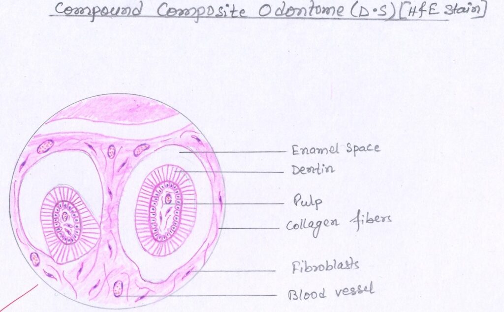
- Normal-appearing enamel or enamel matrix, dentin, pulp tissue and cementum which may or may not exhibit a normal relation to one another
- If the morphologic resemblance to teeth does exist, the structures are usually single-rooted
- The connective tissue capsule around the odontoma is similar in all respects to the follicle surrounding a normal tooth
- Compound Composite Odontoma
- It recapitulates the organization of a normal tooth (the denticles are embedded in fibrous connective tissue, and have a fibrous capsule)
- Complex Composite Odontoma
- Appears as disorganized mass of hard odontogenic tissues, but frequently with a radial pattern. The pulp is usually finely branched so that the mass is perforated, like a sponge, by small branches of pulp
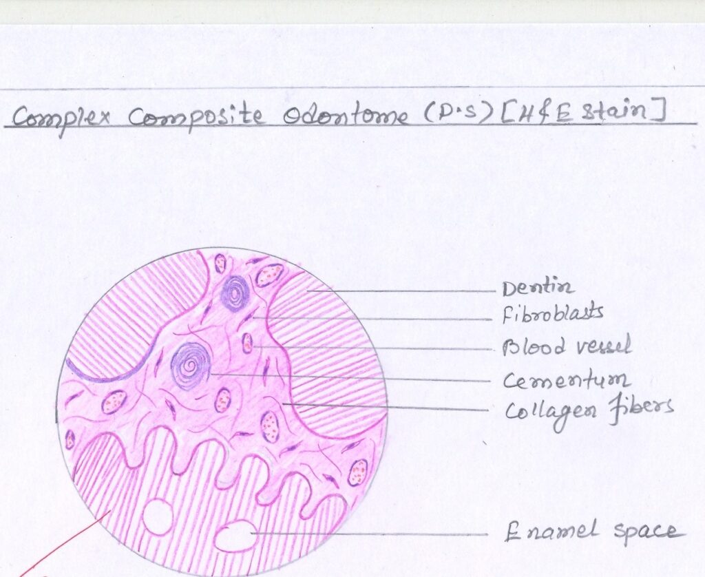
- One additional interesting feature is the presence of ‘ghost’ cells in odontomas. These are the same cells described in the calcifying odontogenic cyst (20% of cases according to a study). No significance attached to the presence of these cells regarding prognosis or treatment of the odontoma.
TREATMENT & PROGNOSIS
- Surgical removal
- No expectancy of recurrence
REFERENCES
- Shafer’s Textbook Of Oral Pathology
- Neville – Oral & Maxillofacial Pathology
Thank you ma’am!
Most welcome
Pretty nice post. I just stumbled upon your blog and
wanted to say that I’ve really enjoyed surfing around
your blog posts. In any case I will be subscribing to your
feed and I hope you write again soon!